
Bridging external fixator (temporary)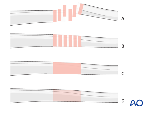 1. Note on illustrationsThroughout this treatment option illustrations of generic fracture patterns are shown, as four different types: A) Unreduced fractureB) Reduced fractureC) Fracture reduced and fixed provisionallyD) Fracture fixed definitively
2. Principles of joint-bridging external fixationA joint-bridging external fixator is fixed to the femur and the tibia while the fracture zone is left virtually untouched (it is bridged). Provisional reduction is achieved with distraction. When external fixation is used to bridge the knee joint it is always only for temporary fixation. Prolonged immobilization will lead to difficulties with knee mobility. It is rapidly applied without need for intraoperative x-rays and can be adjusted later. Details of external fixation are described in the Specific considerations for the knee are given below.
3. Patient preparationThis procedure is normally performed with the patient in a
4. Pin insertion (femur and tibial shaft)Pin placementFor safe pin placement make use of the Any pin placed near a joint should be a minimum of 14 mm away from the joint line to prevent joint sepsis.
Soft-tissue dissectionIn the femur, blunt dissection of the soft tissues and the use of small Langenbeck retractors will minimize muscular damage. Using a straight clamp, prepare a channel for the insertion of the pin.
Tibial pin placementDrilling a hole in the thick tibial crest may be associated with excessive heat generation and there is a risk the drill bit may slip medially or laterally damaging the soft tissues. As the anteromedial tibial wall provides adequate thickness for the placement of pins, this trajectory is preferable. A trajectory angle (relative to the sagittal plane) of 20-60° for the proximal fragment and of 30-90° for the distal fragment is recommended.
Alternatively, in order to avoid the frame catching on the opposite leg, the pins may be placed more anteriorly. The drill bit is started with the tip just medial to the anterior crest, and with the drill bit perpendicular to the anteromedial surface (A). As the drill bit starts to penetrate the surface, the drill is gradually moved more anteriorly until the drill bit is in the desired plane (B). This should prevent the tip from sliding down the medial or lateral surface.
5. Frame construction / reduction and fixation (knee)Teaching videoAO teaching video: Femur--Tibia Articular Fracture Large External Fixator: Knee-bridging Modular Frame Frame assemblyPearl: oblique distal tubeAngle the rod over the tibia such that it is attached to the pins one on the lateral and one on the medial side. This results in a larger window over the condyle which can be beneficial for later (minimally invasive) surgery if it is necessary to maintain the external fixator during definitive osteosynthesis.
Reduction and fixationUsing the partial frames as handles, manually reduce the fracture in length, rotation and axis. Restore length with a bolster behind the knee to give slight flexion. It should be understood that a perfect reduction will not be obtained.
6. Aftercare following joint-bridging external fixationCompartment syndrome and nerve injuryClose monitoring of the tibial compartments should be carried out especially during the first 48 hours after surgery to rule out compartment syndrome. The neurovascular status of the extremity must be carefully monitored. Impaired blood supply or developing neurological loss must be investigated as an emergency and dealt with expediently. Pin-site careVarious aftercare protocols to prevent pin track infection have been established by experts worldwide. Therefore, no standard protocol for pin-site care can be stated here. Nevertheless, the following points are recommended: The aftercare should follow the same protocol until removal of the external fixator. The pin-insertion sites should be kept clean. Any crusts or exudates should be removed. The pins may be cleaned with saline and/or disinfectant solution/alcohol. The frequency of cleaning depends on the circumstances and varies from daily to weekly but should be done in moderation. No ointments or antibiotic solutions are recommended for routine pin-site care. Dressings are not usually necessary once wound drainage has ceased. Pin-insertion sites need not be protected for showering or bathing with clean water. The patient or the carer should learn and apply the cleaning routine. Pin loosening or pin tract infectionIn case of pin loosening or pin tract infection, the following steps need to be taken: Remove all involved pins and place new pins in a healthy location. Debride the pin sites in the operating theater, using curettage and irrigation. Take specimens for a microbiological study to guide appropriate antibiotic treatment if necessary. Before changing to a definitive internal fixation an infected pin tract needs to heal. Otherwise infection will result. Pin loosening or pin track infectionIn case of pin loosening or pin track infection, the following steps need to be taken: Remove all involved pins and place new pins in a healthy location. Debride the pin sites in the operating theater, using curettage and irrigation. Take specimens for a microbiological study to guide appropriate antibiotic treatment if necessary. Before changing to a definitive internal fixation an infected pin track needs to heal. Otherwise infection will result. MobilizationIn order to avoid severe knee stiffness it is important to restart knee movement as soon as possible. If a joint spanning fixator has been applied because of intraarticular fractures these should be reduced and stabilized within the first few days after the injury if possible. Depending on the fracture pattern and soft-tissue injuries, it may be preferable to perform a limited fixation of the articular fragments and convert the external fixator to a non-spanning configuration (see modular external fixation with large pins for extraarticular metaphyseal fractures). Weight bearingWeight bearing is usually restricted following fixation of intraarticular fractures. Follow upSee patient 7-10 days after surgery for a wound check. X-rays are taken to check the reduction.
|


Shanghai Carefix Medical Instrument Co., Ltd.China
沪ICP备16042301号-1 (沪)-非经营性-2016-0122 沪食药监械生产许20101725号
